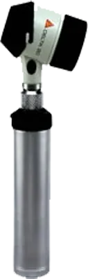
Dermoscopy is the examination of the skin using a hand held skin microscopy system. A Dermoscope used by an experienced Skin Cancer Doctor can make it easier to diagnose melanoma.
Dermoscopy requires a specialized medical instrument with powerful magnifying lens. This allows examination of skin structures and patterns.
The Skin and Mole Clinic is fully equipped with both hand held Dermoscopes and Digital Video Dermoscopy (Molemax).
What Are the Doctors Looking For?
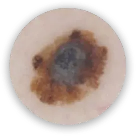
Each pigmented skin lesion can be evaluated in terms of colours and structures.
Colours found in pigmented skin lesions include:
Black, Dark Brown, Light Brown, Red, Blue/Grey, and white.
Australia and New Zealand have the highest rates of melanoma in the world.
In Australia, 1 in 14 males and 1 in 22 females will develop invasive melanoma in their lifetime.
Australia and New Zealand have the highest rates of melanoma in the world.
In Australia, 1 in 14 males and 1 in 22 females will develop invasive melanoma in their lifetime.
Structural Analysis of skin lesions includes:
Symmetry or asymmetry
Homogeny (sameness) or heterogeny (structural differences within the lesion)
A Blue Grey Pigmentation (Highly Senistive for Melanoma)
Distribution of the Pigment Network: Prominent vs Regular, Peripheral and Central black/brown dots and globules